 Living organisms are equipped with 5 senses
that allow us to perceive and interpret our surroundings, with sight being one
of the most vital of these senses. A set of eyes enable us to see the world
around us by converting light waves into electrical impulses that the brain can
perceive. But the eyes are much more than that. Not only do they allow us to connect to the external world through vision, they also project the inner emotions of
people to the external world! The eyes are a complex organ made of intricate
structures and layers, which you will learn more about in this blog.
Living organisms are equipped with 5 senses
that allow us to perceive and interpret our surroundings, with sight being one
of the most vital of these senses. A set of eyes enable us to see the world
around us by converting light waves into electrical impulses that the brain can
perceive. But the eyes are much more than that. Not only do they allow us to connect to the external world through vision, they also project the inner emotions of
people to the external world! The eyes are a complex organ made of intricate
structures and layers, which you will learn more about in this blog.
Origin and Evolution
Most organisms depend on light in some way or
another. Even the more primitive species required and responded to light as an
energy source: plants turn their leaves to face the sun; some algae and other
microorganisms swim towards or away from light. The eyes evolved from simple light
absorbing structures such as chlorophyll. The first eyes, called eye spots, are
thought to have been clusters of photosensitive cells within pigmented pits
that provided directional information to organisms. These pits randomly became
deeper, producing sharper images. Subsequently, the opening narrowed producing
pinhole eyes. The greatest advance in
eyes came when organisms gained the ability to produce images from the light
they collected. These image producing eyes are of two types; camera eyes and
compound eyes4,7.

Source: http://news.nationalgeographic.com/content/dam/news/rights-exempt/nat-geo-staff-graphics-illustrations/2016/01/Evolution_Eyes/eye_evolution_645.jpg?06
Source: http://news.nationalgeographic.com/content/dam/news/rights-exempt/nat-geo-staff-graphics-illustrations/2016/01/Evolution_Eyes/eye_evolution_645.jpg?06
Structure and Funtion of the eye
 |
| http://healthfavo.com/wp-content/uploads/2013/08/human-eye-diagram-labeled.jpg |
Cornea: A transparent, curved layer that acts
as the first lens of the eye and lets light enter
Sclera: A thin opaque layer of connective tissue that
acts as a supporting wall, lending rigidity. It begins at the end of the
cornea1.
Pupil: The dark coloured aperture that allows light to enter into the
posterior section of the eye1.
Iris: a pigmented circular muscle that surrounds and controls the size of the
pupil
Lens: is a transparent body that focuses the light onto the retina
Retina: The innermost layer of the eye containing photosensitive cells that
respond to light1.
Chambers
of fluid: There
are three fluid filled chambers in the eye; the Anterior Chamber (between
cornea and iris) the Posterior chamber (between iris and lens) and the Vitreous chamber (between
lens and retina) which provide structural integrity to the eye and prevent it
from collapsing1.
Choroid: A vascular layer that sits between the retina and the sclera and nourishes the surrounding layers1.
Choroid: A vascular layer that sits between the retina and the sclera and nourishes the surrounding layers1.
Pathwhay of Light in the Eye
 |
| https://aos.iacpublishinglabs.com/question/aq/700px-394px/NULL_38229003d3d8213b.jpg?domain=cx.aos.ask.com |
The eye itself is a complex structure made up of many layers. the
external surface of the eye is made of a thin layer of connective tissue, known
as the sclera. This opaque white layer gives the eyeball its white colour and forms
part of the supporting wall, thereby protecting the eye1.
At the anterior end of the eyeball is a transparent, curved layer known as the cornea, which serves as the first lens of the eye as it helps to focus light as it initially enters the eye. The pupil is the dark coloured aperture that allows light to enter into the posterior section of the eye. It is black due to the presence of absorbing pigments. The coloured layer around the pupil is known as the iris. This is a pigmented circular muscle that controls the size of the pupil via a process known as the pupillary reflex. In a bright environment, the iris causes the pupil to constrict, allowing very little light to enter. In low light areas, the iris causes the pupil to dilate, allowing more light to enter the eye1.
Behind the iris is a transparent body known as the lens. The lens serves to focus the images onto the retina. For objects close by, the lens becomes thicker by the relaxation of ciliary muscles. To focus on objects at a distance, the ciliary muscles contract, causing the lens to elongate. Once the light has focused, it hits the retina, a layer which contains photoreceptors. These are the light sensitive cells present in the eye that respond to the light. The macula is the most sensitive region of the retina as it has the highest density of photoreceptors. Light is most often focused onto this region as it produces the most detailed images due to the abundance of photoreceptors. The photoreceptors are connected to nerve fibres, which bundle up to form the optic nerve. Once light hits the photoreceptors, it is converted into an electrical signal that is carried to the brain by the optical nerve1.
The sclera is a dense layer of connective tissue that extends from the cornea. The corneal limbus is the reigon of junction between the two. Like the cornea, the sclera is composed of dense bundles of collagen fibres with flattened fibroblasts scattered in between. Elastic fibrils are also present within the sclera. Toward the posterior end, near the optic nerve, the sclera becomes a thin
fenestrated membrane known as the lamina cribosa. The sclera fuses into the
dura of the the optic nerve2,3.
At the anterior end of the eyeball is a transparent, curved layer known as the cornea, which serves as the first lens of the eye as it helps to focus light as it initially enters the eye. The pupil is the dark coloured aperture that allows light to enter into the posterior section of the eye. It is black due to the presence of absorbing pigments. The coloured layer around the pupil is known as the iris. This is a pigmented circular muscle that controls the size of the pupil via a process known as the pupillary reflex. In a bright environment, the iris causes the pupil to constrict, allowing very little light to enter. In low light areas, the iris causes the pupil to dilate, allowing more light to enter the eye1.
Behind the iris is a transparent body known as the lens. The lens serves to focus the images onto the retina. For objects close by, the lens becomes thicker by the relaxation of ciliary muscles. To focus on objects at a distance, the ciliary muscles contract, causing the lens to elongate. Once the light has focused, it hits the retina, a layer which contains photoreceptors. These are the light sensitive cells present in the eye that respond to the light. The macula is the most sensitive region of the retina as it has the highest density of photoreceptors. Light is most often focused onto this region as it produces the most detailed images due to the abundance of photoreceptors. The photoreceptors are connected to nerve fibres, which bundle up to form the optic nerve. Once light hits the photoreceptors, it is converted into an electrical signal that is carried to the brain by the optical nerve1.
Histology
They eye is
composed of three main layers; the outermost layer is called the fibrous layer
and is composed of the cornea at the and the sclera. The middle layer is a
vascular layer that includes the choroid, ciliary body and the iris. The
innermost layer is known as the neural layer and this consists of the retina2.

Fibrous Layer
The external
surface of the cornea is covered with non-keratinized stratified squamous
epithelium, with columnar cells sitting at the basement membrane. Below the
epithelial layer is an acellular region of collagen bundles known as Bowman’s
membrane2. The stroma (also known as substantia propria) is the body of the
cornea, composed of laminae of collagen fibrils. Some elastic fibrils are also
present within the lamina. Flattened fibroblasts as well as some macrophages
and leukocytes can be found between the lamina. Below the stroma is the
acellular basal lamina of the posterior epithelium layer composed of collagen.
The posterioir corneal epithelium is composed of a single layer of cuboidal
cells.
 |
| https://blogger.googleusercontent.com/img/b/R29vZ2xl/AVvXsEjF3H5fE6ZFVF2yHclJJbQDTXzZJbcpBNjD3Eg7VRDnn3RIB6vD2DeWX6I_J-rdnxdk7lvxOdtwFCPQZQ_PtaT_le6Wyhx1iqIzxge193FmBbXFMOWOjME3GBIID0Ne6Kx4zUgLFy9eeXU/s1600/cornea.png |
 |
| Lamina cribosa |
Vascular Layer
The vascular
layer is composed of the iris, ciliary body and the choroid.
 |
| http://clinicalgate.com/wp-content/uploads/2015/03/B9781437719260100037_f03-05-9781437719260.jpg |
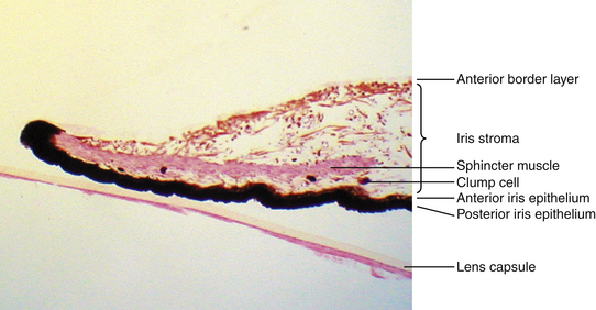
The iris is composed of four layers: (1) the anterior border densely lined with
pigmented or non-pigmented cells. (2) The stroma, composed of loose vascular
pigmented connective tissue with melanocytes (pigmented cells that are absent
in blue eyes), fibroblasts and phagocytes. (3) The dilator muscle layer (4) Posterior
epithelium, made of two layers of pigmented epithelium. At the inner border of
the iris, where the pupil is formed, lie the smooth muscles of the sphincter
muscle of the iris2,3.
The choroid
is a richly vascularized region also composed of four distinct layers (in order
from the sclera to the retina): (1)The suprachoroid layer is similar to the
sclera and is composed of lamellae of collagen fibres with scattered elastic fibres
and interspersed fibroblasts, macrophages and melanocytes. (2) The vascular
layer is composed of large muscular vessels. The stroma of the vascular layer
is composed of loose connective tissue withy interspersed melanocytes. (3) The
choriocapillary layer has a stroma made of fine collagen and elastic fibres,
with large capillaries present within the stroma. (4) The Lamina Vitrea layer
is the basal lamina of the pigment layer of the retina and i composed of two
layers of collagen fibres with elastic fibres in between2,3.
http://www.intechopen.com/source/html/37889/media/image1.jpeg
 |
| 1: Sclera; 2: Suprachoroid; 3: Large-sized- vessel layer (Haller´s Layer); 4: Medium-sized-vessels; 5: Choriocapillaris; 6: Bruch´s membrane; 7: retinal pigment epithelium. |
http://www.intechopen.com/source/html/37889/media/image1.jpeg
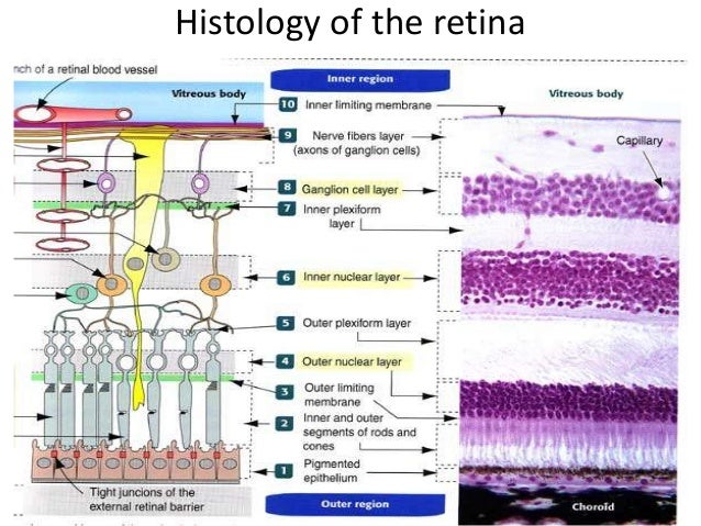
The neural
layer is the retinal layer, composed of nine distinct regions (starting
externally from the choroid):
(1) The outermost layer is the pigment epithelium, formed of a single layer of cuboidal cells that contain pigment in the apical region. Extending into the adjacent layer are processes of pigment granules. The pigment epithelium is separated from the photoreceptor layers by the subretinal space.
(1) The outermost layer is the pigment epithelium, formed of a single layer of cuboidal cells that contain pigment in the apical region. Extending into the adjacent layer are processes of pigment granules. The pigment epithelium is separated from the photoreceptor layers by the subretinal space.
(2) The second
layer is composed of photoreceptor cells: thin rod cells and thicker cone cells.
(3) The outer
limiting membrane lies beneath the layer of rods and cones. The processes of
neural cells known as Muller’s cells can be found within this layer.
(4)The outer
nuclear layer contains the nuceli of rods and cones as well as the outer processes
of the Muller cells.
(5) The outer
plexiform layer is where the prhotorecptive cells from synapses with the the
processes of neural cell cells, the bodies of which are present in the
subsequent layer.
(6) The inner
nucler layer contains the nuclei and cell bodies of three types of neural cells: amacrine cells,
horizontal cells and bipolar cells as well as the nuclei of the Muller cells.
This is where the intial processing of the senscory input occurs.
(7)The inner
plexiform layer is where the axons of the bipolar cells (whose bodies are in
the inner nuclear layer), synapse with the dendrites of ganglion cells, whose
bodies are present within the subsequent layer (Layer 8).
(8) The
ganglion cell layer houses the large multipolar ganglionic cell bodies as well
as scattered neuroglia. These cells are more abundant near the fovea, but less
so near the periphery of the retina.
(9) The layer
of optic nerve fibres contains the axons of the ganglion cells from the
ganglionic cell layer and the inner processes of the Muller’s cells. The axons
travel towards the optic disc, where they gather and converge, forming the optic nerve.(10) The
inner limiting membrane is the basal lamina composed of the terminations of the
inner fibres of Muller’s cells2,3,6.
The following video provides a general summary of the pathway of light in the eye and of the retinal layers:
(10) The
inner limiting membrane is the basal lamina composed of the terminations of the
inner fibres of Muller’s cells.
The following video provides a general summary of the pathway of light in the eye and of the retinal layers:
Pathology
Age related macular degeneration
A major cause of blindness for seniors in
North America, macular degeneration is a disease caused by defects in Burch’s
membrane. The membrane becomes fenestrated and thickens, cuasing blood vessels
to grow through into the retina, which can leak fluid into the retina causing
atrophy of the pigment epithelium. If the blood vessels leak, a hemorrhage can
occur, effecting the quality of vision. AMD
can also be of the dry form wherein drusen, dried lipids and fatty protein, can
accumulate between the pigment epithelium and Burch’s membrane6.
Diabetic retinopathy
Diabetic retinopathy is a diabetic eye
disease that can affect people with diabetes. High blood sugar can damage the
vessels present in the small blood vessels at the back of the eye, causing hemorrhaging,
which in turn lead to swelling and decreased vision. Another way diabetic
retinopathy can deteriorate vision is by causing swelling of the macula causing
macular edema, which may cause blindness6.
References:
((1) Kolb, H., Nelson, R., Fernandez, E., & Jones,
B. (Eds.). (n.d.). Webvision. Retrieved October 25, 2016, from
http://webvision.med.utah.edu/
(2) Hinrichsen, C. (1997). Organ histology: A
student's guide. Singapore: World scientific.
(3) Eroschenko, V. P., & Fiore, M. S.
H. (1996). Di Fiore's atlas of histology with functional correlations.
Baltimore: Williams & Wilkins.
(4) Markgraf, B. (n.d.). Insect Compound Eye vs. Human
Eye | The Classroom | Synonym. Retrieved October 25, 2016, from
http://classroom.synonym.com/insect-compound-eye-vs-human-eye-22657.html
(5) Ophthalmic Pathology: Altas Web Site. (n.d.).
Retrieved October 25, 2016, from http://www.ouhsc.edu/ocupath/
(6) Slomianka, L. (2009, August 5). Blu Histology -The
Eye. Retrieved October 25, 2016, from
http://www.lab.anhb.uwa.edu.au/mb140/corepages/eye/eye.htm#retina
(7) Yong, E. (2016, January 14). Inside the Eye:
Nature's Most Exquisite Creation. Retrieved October 25, 2016, from
http://ngm.nationalgeographic.com/2016/02/evolution-of-eyes-text